1️⃣ Basic Details Section
Usually at the top of the report.
- Patient name / age
- LMP (Last Menstrual Period)
- GA (Gestational Age)
→ Calculated by LMP or ultrasound measurements
- EDD (Estimated Due Date)
👉 Small variation between LMP GA and scan GA is common (±5–7 days in early pregnancy).
2️⃣ Pregnancy Location & Viability
✔️ Intrauterine pregnancy (IUP)
Confirms pregnancy is inside uterus (rules out ectopic).
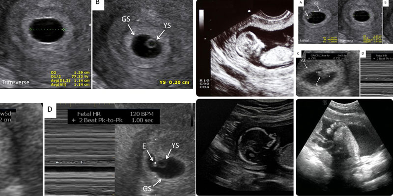
✔️ Gestational sac (GS)
Seen from ~4.5–5 weeks.
✔️ Yolk sac
First structure confirming viability.
✔️ Fetal pole + cardiac activity
Normal ranges:
- 6–7 weeks → ~110–160 bpm
- Later pregnancy → 120–160 bpm
3️⃣ Gestational Age Measurements
📏 First trimester parameter
CRL (Crown-Rump Length)
👉 Most accurate dating measurement (up to 13+6 weeks)
📏 Second & third trimester biometry
Standard fetal measurements:
- BPD — Biparietal diameter (head width)
- HC — Head circumference
- AC — Abdominal circumference
- FL — Femur length
👉 These calculate:
- GA (by scan)
- EFW (Estimated Fetal Weight)
✔️ Values are plotted against percentile charts.
4️⃣ Fetal Anatomy Assessment
(Usually detailed in anomaly scan ~18–22 weeks)
- Brain structures
- Face (lip/palate)
- Spine
- Heart (4-chamber view, outflow tracts)
- Abdomen (stomach, kidneys, bladder)
- Limbs
👉 Report often states “No obvious structural abnormality detected.”
5️⃣ Placenta Evaluation
- Location → Anterior / Posterior / Fundal
- Grade → Maturity (0–III)
- Distance from os → Important if placenta is low-lying
6️⃣ Amniotic Fluid
- AFI (Amniotic Fluid Index)
Normal ≈ 8–24 cm
OR
- SDP (Single Deepest Pocket)
Normal ≈ 2–8 cm
7️⃣ Cervix & Maternal Structures
- Cervical Length
👉 >30 mm → Normal length
👉 <25 mm → Increased risk of preterm birth
- Uterus & Adnexa
→ fibroids, ovarian cysts
8️⃣ Doppler Study (if done)
- Umbilical Artery
- MCA (Middle Cerebral Artery)
- Uterine Artery
Used for:
- Fetal Growth Restriction (FGR)
- Preeclampsia
- High-risk pregnancies
🧠 How to Interpret Overall Impression
The “Impression” or “Conclusion” section summarizes the key findings of the ultrasound report.
Examples:
- Single live intrauterine pregnancy of ___ weeks
- Fetal growth appropriate for gestational age
- Low-lying placenta
- Mild oligohydramnios
👉 Always correlate the ultrasound findings with the clinical context and your doctor's assessment.
⚠️ Red Flags to Look For
- GA (Gestational Age) mismatch > 2 weeks
- Absent cardiac activity
- EFW (Estimated Fetal Weight) < 10th percentile
- AFI (Amniotic Fluid Index) low or high
- Placenta covering the internal os
- Short cervix
📝 Quick Interpretation Flow
- Confirm viability
- Check GA vs LMP
- Assess growth parameters
- Look at placenta & AFI
- Read the impression
✅ Bottom Line
A pregnancy ultrasound report answers three core questions:
👉 Is the baby alive?
👉 Is growth appropriate?
👉 Is the environment (placenta + fluid + cervix) normal?
❓ Other Frequently Asked Questions on Ultrasound in Pregnancy
1️⃣ How many ultrasounds are done in a normal pregnancy?
Typically 3–4 routine scans:
- Early viability scan (6–8 weeks) – usually done transvaginally
- NT scan (11–13+6 weeks)
- Anomaly scan (18–22 weeks)
- Growth scan (32–36 weeks, if needed)
👉 High-risk pregnancies may require additional scans.
2️⃣ Is ultrasound safe during pregnancy?
Yes ✅
Ultrasound uses sound waves (not radiation) and is considered safe for both mother and baby when performed for medical reasons.
3️⃣ Which ultrasound is most important?
👉 The anomaly scan (18–22 weeks)
Because it evaluates the baby's organs and helps detect structural abnormalities.
4️⃣ Why is ultrasound dating sometimes different from LMP?
This can happen because:
- Ovulation timing may vary
- Menstrual cycle length may not be exactly 28 days
👉 First-trimester CRL dating is the most accurate method.
5️⃣ Can ultrasound detect all birth defects?
No ❌
Ultrasound can detect many structural abnormalities, but not all genetic or minor conditions.
Detection depends on:
- Gestational age
- Fetal position
- Equipment quality
- Examiner expertise
6️⃣ What is a growth scan?
A scan performed during the third trimester to assess:
- Baby's weight
- Amniotic fluid
- Placenta
- Doppler studies (if required)
👉 Used to monitor fetal growth and wellbeing.
7️⃣ Why is my due date changed after a scan?
If early ultrasound dating differs significantly from LMP, the EDD may be revised because early pregnancy measurements are generally more reliable.
8️⃣ Can ultrasound determine baby's position?
Yes 👍
Especially in the third trimester, ultrasound can determine whether the baby is:
- Head-down (Cephalic)
- Breech
- Transverse
9️⃣ What does “single live intrauterine pregnancy” mean?
It simply means:
- ✔️ One baby
- ✔️ Located inside the uterus
- ✔️ Heartbeat is present
A normal and reassuring finding.
🔟 Do I need a full bladder for pregnancy ultrasound?
- Early pregnancy (Transabdominal scan) → Often yes
- Later pregnancy → Usually not required
👉 What does AFI mean in pregnancy ultrasound?
AFI (Amniotic Fluid Index) measures the amount of fluid surrounding the baby and helps assess fetal wellbeing.
❓ How long does a pregnancy ultrasound take?
- Routine scans usually take 15–30 minutes
- Detailed anomaly scans may take 30–45 minutes
👉 Extra time may be needed if the baby's position makes imaging difficult.
❓ Does a pregnancy ultrasound hurt?
No — it is generally painless ✅
- Transabdominal scan: Mild pressure from the probe may be felt
- Transvaginal scan: Slight discomfort may occur but is usually not painful
There are typically no after-effects, and normal activities can be resumed immediately.
❓ Why is a pregnancy ultrasound sometimes done transvaginally?
A transvaginal ultrasound (TVS) provides a clearer and more detailed view during early pregnancy.
Why TVS may be preferred:
- The probe is closer to the uterus, resulting in better image resolution
- Helps detect very early pregnancy (4–9 weeks)
- Better for confirming early cardiac activity
- Useful when abdominal scan visibility is limited (retroverted uterus, obesity, empty bladder, etc.)
👉 TVS is performed only when medically indicated and is considered safe during pregnancy.